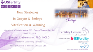


Session 170: Jamais Vu

Donate
At the International IVF Initiative, we are committed to providing free access to our educational sessions, webinars, and resources for professionals and individuals passionate about advancing reproductive medicine. We believe that cost should never be a barrier to knowledge and collaboration. By contributing, you’re ensuring that valuable educational resources, expert insights, and collaborative opportunities remain open to all without financial barriers. Together, we can continue to foster a global community dedicated to innovation and excellence in the field of IVF.
Your Donation
Thank you!
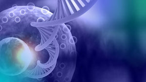
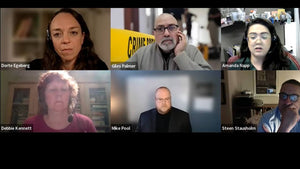
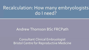
Session 170: Jamais Vu: Cell imaging webinar
Tuesday, 14th April 2026, at 3pm EST/UK/9pm CET.
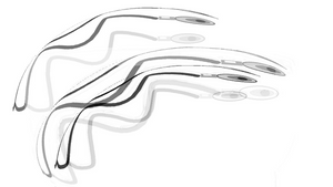
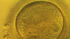
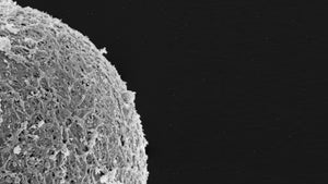
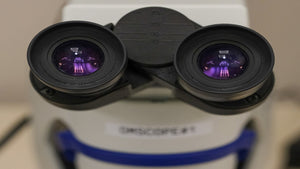
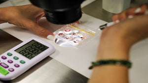
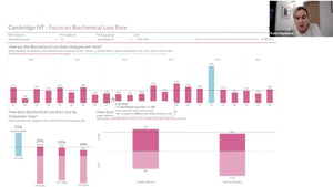
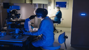
Image courtesy of The Plachta Lab
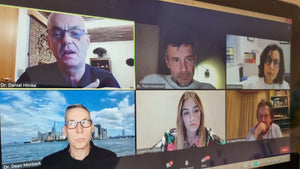
Moderators:
Dr. Emna Ouni, Dr. Debbie Monjean and Dr. Gerardo Mendizabal-Ruiz
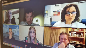
Presenters:
Dr. Emna Ouni: Label-free imaging in IVF: a game of light and shadows
Dr. Nicolas Plachta: Imaging how the embryo forms in real time
Dr. Erick Vargas-Ordaz & Dr. Fab Horta Nunez : Metabolic imaging of early embryos using a light-sheet on-a-chip device
Q and A
Synopsis
The webinar *Session 170: Jamais Vu: Cell Imaging* took place on Tuesday, 14th April 2026, bringing together leading experts to explore emerging imaging technologies in embryology and reproductive science. Moderated by Dr. Emna Ouni, Dr. Debbie Monjean, and Dr. Gerardo Mendizabal-Ruiz, the session focused on how innovative, non-invasive imaging approaches were reshaping the understanding of early embryo development.
The session opened with a presentation by Dr. Emna Ouni, who discussed the application of label-free imaging in IVF. She described how this approach leveraged intrinsic optical properties of cells—such as light scattering, refractive index variations, and endogenous contrast—to visualize embryo structures without the need for fluorescent labels or dyes. Her talk emphasized how this “game of light and shadows” enabled clinicians and researchers to assess embryo quality in a more physiological and non-disruptive manner. She highlighted the potential of label-free techniques to improve embryo selection by preserving viability while still extracting rich morphological and dynamic information.
Following this, Dr. Nicolas Plachta presented on real-time imaging of embryo formation. He outlined how advanced live-cell imaging technologies had allowed researchers to observe developmental processes as they unfolded, capturing cell fate decisions, spatial organization, and lineage specification in unprecedented detail. His work illustrated how time-resolved imaging could reveal the dynamic choreography of early embryogenesis, moving beyond static snapshots to a continuous understanding of development. He also addressed the computational challenges associated with analyzing such complex datasets and the importance of integrating imaging with quantitative modelling.
The session then transitioned to a joint presentation by Dr. Erick Vargas-Ordaz and Dr. Fab Horta Nunez, who introduced a novel approach to metabolic imaging using a light-sheet on-a-chip device. They explained how this platform combined the advantages of light-sheet microscopy—such as reduced phototoxicity and rapid volumetric imaging—with microfluidic technology to create a controlled environment for embryo culture and observation. Their work focused on measuring metabolic activity in early embryos, providing insights into cellular energy states and developmental potential. The presenters emphasized how this technology could enable high-throughput, non-invasive metabolic profiling, potentially offering new biomarkers for embryo viability.
The webinar concluded with a Q&A session, during which participants engaged with the speakers on topics such as clinical translation, scalability of imaging technologies, and ethical considerations in embryo research. Discussions also touched on the integration of artificial intelligence for image analysis and the future role of multimodal imaging in IVF laboratories.
Overall, the session underscored a shift toward non-invasive, high-resolution, and dynamic imaging techniques, highlighting their potential to transform both research and clinical practice in reproductive biology.
Dr. Emna Ouni
Driven by a passion for engineering innovation in human health, Dr. Emna Ouni is an industrial bioengineer whose work bridges biomimetics, advanced imaging, and tissue engineering. Trained at leading institutions including, European research center for Diabetes (CEED), UCLouvain, and Institut Gustave Roussy, she began her scientific journey in 2015 developing 3D models for transplantable artificial pancreas, in Strasbourg, France. Her research evolved toward the matrisome and extracellular matrix as key determinants of biofunctional design. During her PhD, she introduced matrisome-driven approaches to reproductive biology, producing the first imaging-informed blueprint of the human ovarian niche. A two-time successive European grant laureate under the MSCA Horizon Europe program and principal inventor of a patent, she now pioneers high-throughput tumoroid systems and label-free holotomography-compatible 3D models. An invited speaker at flagship imaging events and instructor, she advances precision medicine and next-generation therapeutics.
Dr. Debbie Monjean
Dr Debbie Montjean received her doctoral degree in 2011 (University Paris VI) after she had graduated from a master’s degree in physiology and physiopathology with a specialization in reproductive and developmental biology (University Paris VII). Debbie served as deputy of ESHRE SIG embryology for 4 years Dr Debbie Montjean worked during 9 years as a clinical and research embryologist in France and since 2021, she works as the IVF laboratories director and Scientific Innovation Advisor at Fertilys fertility clinics (Canada).
Dr. Gerardo Mendizabal-Ruiz
Gerardo is a researcher and inventor specializing in automation and in vitro fertilization (IVF), with a strong focus on developing advanced technological systems for reproductive medicine. His work integrates engineering, computer vision, robotics, and AI to design precise, scalable, and reliable solutions. He has experience in applied research, prototyping, and patent development, bridging the gap between scientific theory and real-world implementation. Gerardo is driven by innovation, efficiency, and measurable impact, aiming to create technologies that improve clinical outcomes and expand the possibilities of assisted reproduction
Dr. Nicolas Plachta
Dr. Erick Vargas-Ordaz
He has held research roles at Monash University and the Melbourne Centre of Nanotechnology, focusing on optical system integration and live biological imaging. Dr Vargas-Ordaz is an inventor on patent filings for non-invasive imaging systems and has published in leading journals, with expertise in translating research into scalable biomedical technologies.
Dr. Fab Horta Nunez